Long-Term Survival of a Maxillary Subperiosteal Implant After Blast Injury: A 17-Year Follow-Up Case Report
In depth4:11CCAI
paperi.ai
0:00 / 0:00
Petro Rosiiskyi, Luigi Angelo Vaira
A dental implant endured seventeen years in a severely atrophic upper jaw, and then a high-energy blast injury. The remarkable part is what the implant did not do: it did not become mobile or lose its functional stability.
Subperiosteal implants (SPIs) have re-emerged as a treatment option for the rehabilitation of patients with severe maxillary atrophy, although evidence on their long-term performance remains limited. We report the case of a 53-year-old female patient who underwent maxillary rehabilitation with a custom subperiosteal implant in 2006 due to insufficient bone volume for conventional endosseous implant placement. The implant was fabricated using a direct bone impression technique and designed to achieve mechanical engagement within the anatomical undercuts of the maxilla, providing primary stabilization without extensive screw fixation. The patient maintained satisfactory functional and aesthetic outcomes over a 17-year follow-up period. In 2023, she sustained a high-energy facial blast injury resulting in damage to the prosthetic superstructure. Clinical and radiological evaluation revealed preservation of implant stability, absence of pathological bone resorption, and only a localized intrusion of the implant framework of approximately 2 mm in the anterior region. No implant mobility or functional impairment was observed. The patient was successfully rehabilitated with a new prosthetic restoration without the need for additional surgical intervention. This case highlights the potential long-term stability of subperiosteal implants and provides a rare clinical observation of their behavior under extreme traumatic conditions. Further studies are needed to better understand the biomechanical performance of these systems.
Transcript
A dental implant endured seventeen years in a severely atrophic upper jaw—and then a high-energy blast injury. The remarkable part is what the implant did not do: it did not become mobile or lose its functional stability. Severe maxillary atrophy remains a major challenge in implant rehabilitation, often precluding the use of conventional endosseous implants because of insufficient bone volume.
In such cases, subperiosteal implants, or SPIs, may offer an alternative approach, allowing prosthetic rehabilitation without extensive bone grafting procedures. Computer-aided manufacturing design and additive manufacturing have improved framework precision and reproducibility.
However, long-term clinical data remain limited, particularly regarding implant behavior under extreme mechanical stress or traumatic events. This case involves a maxillary subperiosteal implant with seventeen years of follow-up that maintained structural integrity and clinical function after a high-energy blast injury.
That combination makes the case unusual: it examines long-term performance and behavior after a high-energy blast injury in the same patient. After surgical exposure of the maxilla, a direct impression of the osseous surface was obtained.
Anatomical undercuts were identified and incorporated into the framework design to provide mechanical engagement and primary stabilization through a clip-fit mechanism. The implant was placed fourteen days later and showed stable adaptation without the need for extensive screw fixation.
In panel C, the frontal view shows the continuous anterior row of metal-ceramic teeth, while panel D reveals the same restoration from above together with the removable prosthesis and its visible metal framework. The four-panel sequence therefore documents the transition from the severely compromised preoperative appearance to a completed, integrated prosthetic arrangement.
Following the blast injury, the maxillary prosthetic superstructure was damaged, with fracture and loss of the anterior ceramic component. Clinical examination revealed peri-implant soft tissue inflammation, but no implant mobility or pain under functional loading. In panel B, the occlusal view shows the healed palate with the implant components still distributed around the maxilla, while panel C exposes the underlying metallic framework beneath the removed prosthesis.
Panel D then shows a continuous fixed restoration in place, with the anterior teeth integrated into the final smile. Radiographic evaluation showed preservation of the subperiosteal implant framework, without pathological bone resorption. A localized anterior intrusion of approximately two millimeters was observed, while the distal extensions remained stable.
In this case, the absence of mobility, lack of pathological bone resorption, and successful prosthetic re-intervention without further surgery suggest long-term functional stability. Despite localized anterior intrusion after trauma, the framework remained clinically stable.
The stability may be related to the broad distribution of forces across the implant surface and intimate adaptation to the maxillary bone. Clip-fit engagement within anatomical undercuts may have contributed to mechanical retention, although no causal conclusion can be drawn from a single case.
This report is limited by its single-case design and lack of three-dimensional imaging or biomechanical analysis. Larger studies are needed to clarify the long-term biomechanical behavior of subperiosteal implants and the role of different fixation and engagement strategies.
This single case suggests that a custom subperiosteal implant can remain clinically stable for seventeen years and through extreme trauma, although larger studies are needed before drawing broader biomechanical conclusions.
A derivative work by Paperi · AI-generated script, voice and captions
· pages and figures unaltered
Made with Paperi.
Drop in a research PDF — get a narrated video walkthrough like this one,
with highlights that follow the narration. Free to start.
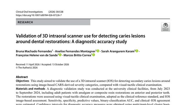
Bruna Machado Fernandes, Anelise Fernandes Montagner, Sarah Arangurem Karam, Françoise Hélène van de Sande, Marcos Britto Corrêa
A filling can look perfectly ordinary while decay is forming at its edge. This study asks whether a three-dimensional dental scan can help find that hidden trouble, without replacing the dentist’s judgment.A dental scanner can produce a remarkably convincing three-dimensional picture, but can that picture actually detect decay around an old filling? This study finds a useful answer: sometimes, but especially as a complement, not a replacement.
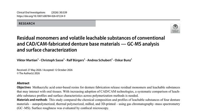
Viktor Martian, Christoph Sasse, Ralf Bürgers, Andrea Schubert, Oskar Bunz
A denture can contain leftover building blocks from its manufacture. But in this study, the material with the most detectable residue did not release more of one key substance than plain control liquid.A denture resin can contain residual monomer without releasing more methyl methacrylate than a negative control. And the 3D-printed material may look chemically different for reasons a single screening method cannot fully reveal.
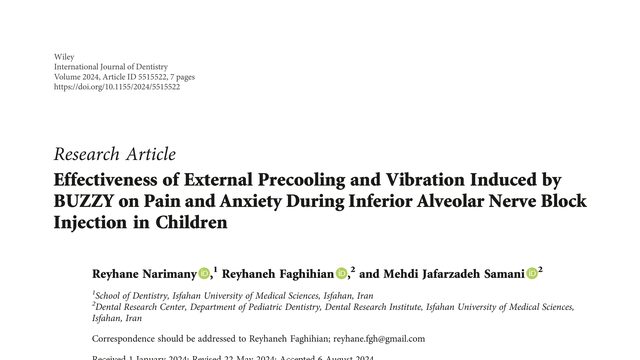
Reyhane Narimany, Reyhaneh Faghihian, Mehdi Jafarzadeh Samani
A small device combines cooling and vibration to make a dental injection less frightening for children. But when it was tested, the reassuring sensation did not clearly reduce either pain or anxiety.A device combining cold and vibration sounds like a simple way to make dental injections easier for children. But in this randomized trial, BUZZY did not significantly reduce pain or anxiety during the injection.
 3:27