Validation of 3D intraoral scanner use for detecting caries lesions around dental restorations: A diagnostic accuracy study
Overview3:27CCAI
paperi.ai
0:00 / 0:00
Bruna Machado Fernandes, Anelise Fernandes Montagner, Sarah Arangurem Karam, Françoise Hélène van de Sande, Marcos Britto Corrêa
A filling can look perfectly ordinary while decay is forming at its edge. This study asks whether a three-dimensional dental scan can help find that hidden trouble, without replacing the dentist’s judgment.
Objectives This study aimed to validate the use of a 3D intraoral scanner (IOS) for detecting secondary caries lesions around restorations using image-based CARS-derived severity categories, compared with visual-tactile clinical examination. Materials and methods A diagnostic validation study was conducted at the university clinical facilities, from July 2023 to September 2024, including adult patients with amalgam or composite resin restorations on anterior and posterior teeth. The restorations were assessed using visual-tactile clinical examination, adopted as the clinical reference standard, and IOS image-based assessment. Sensitivity, specificity, predictive values, binary-classification AUC, and clinical–IOS agreement were estimated. Confidence intervals for diagnostic accuracy measures were obtained using participant-level cluster boot strap resampling. Results A total of 299 restorations in 41 patients were assessed. IOS sensitivity varied, with 75% (95% CI: 66.2–82.0) at the initial lesion threshold, 40% (95% CI: 20.8–57.7) at the moderate lesion threshold, and 57.1% (95% CI: 33.3–100.0) at the severe lesion threshold. Specificity was consistently high, with 84.1% (95% CI: 78.7–89.4), 97.4% (95% CI: 95.6–99.2), and 99.7% (95% CI: 98.7–99.7) at the initial, moderate, and severe lesion thresholds, respectively. Binary-classification AUC values were 0.79 (95% CI: 0.75–0.84), 0.69 (95% CI: 0.58–0.78), and 0.78 (95% CI: 0.66–1.00) at the initial, moderate, and severe lesion thresholds, respectively. Positive predictive value was highest at the severe lesion threshold (80%), while the negative predictive value was especially high at this threshold (99%). Exact clinical-IOS agreement was 77.9%, whereas linearly weighted observed agreement was 91.1%, with a linearly weighted kappa coefficient of 0.53. Conclusions The 3D intraoral scanner is a complementary tool for assessing secondary caries lesions, showing high specific ity across lesion thresholds. Sensitivity varied according to lesion severity and was less precise at the severe threshold due to the small number of severe lesions. Future research should increase sample sizes and explore combining IOS with other diagnostic methods to improve detection, especially for moderate lesions and higher caries prevalence. Clinical Relevance Conventional IOS images may assist in documenting and assessing visible changes around restorations. Clinical interpretation should consider the observed sensitivity limitations and incorporate visual-tactile examination and radiographic assessment when indicated. Keywords Secondary caries · Intraoral scanner · CARS criteria · Diagnostic accuracy · Sensitivity · Specificity
Transcript
A filling can look perfectly ordinary while decay is forming at its edge. This study asks whether a three-dimensional dental scan can help find that hidden trouble—without replacing the dentist’s judgment. Decay around an existing restoration is one of the main reasons a restoration is repaired or replaced, regardless of the material used.
These lesions develop around an existing restoration when marginal failures allow bacteria to penetrate and initiate demineralization of the tooth structure. If they go unnoticed and remain untreated, these lesions may progress over time and lead to further damage of the tooth structure.
These close-up scans capture how restored back teeth can look very different from one another, from smooth, tooth-coloured surfaces to darkened or visibly worn areas. That variety is why judging disease around a restoration from images alone is not straightforward.
Finding decay around a restoration is difficult because the restoration itself can hide what is happening at its edge. The usual approach combines a hands-on examination with an x-ray. The hands-on examination checks visible surfaces, while an x-ray can show decay between teeth, how deep it is, and how close it is to the tooth’s nerve.
The study tested whether a three-dimensional intraoral scanner could detect decay around restorations, using image-based severity categories and comparing its results with a visual-tactile clinical examination. The expectation was that the scans might perform about as well as a hands-on examination, while their performance could change depending on how severe the decay was.
The scanner used ordinary white light to create three-dimensional models that matched the natural color of the teeth, with the teeth kept moist during scanning. A scan counted as adequate when the software captured enough detail of the teeth being examined.
The participants were adults between eighteen and sixty years old; in total, the sample included thirty-four women and seven men. The restorations included both front and back teeth, with forty-six located in anterior teeth and two hundred fifty-three in posterior teeth.
Most of the restorations examined were in back teeth, rather than front teeth. The scan found seventy-five percent of the earliest lesions, but only forty percent of moderate lesions and fifty-seven point one percent of severe lesions.
Its ability to correctly identify restorations without lesions stayed high: eighty-four point one percent for the earliest threshold, ninety-seven point four percent for moderate lesions, and ninety-nine point seven percent for severe lesions. Overall, the scan performed better when the earliest and severe lesion thresholds were considered.
Its ability to rule out lesions stayed high across severity levels, while its positive predictive value was highest for severe lesions. Because severe lesions often lead to repair or replacement, the scan may be useful as an extra way to assess restored teeth, alongside clinical judgment and, when needed, an x-ray.
The scan was good at ruling out serious decay around restorations, but less reliable for catching every lesion, especially moderate ones. It is best used as an extra view alongside examination and, when needed, radiography.
A derivative work by Paperi · AI-generated script, voice and captions
· pages and figures unaltered
Made with Paperi.
Drop in a research PDF — get a narrated video walkthrough like this one,
with highlights that follow the narration. Free to start.
Viktor Martian, Christoph Sasse, Ralf Bürgers, Andrea Schubert, Oskar Bunz
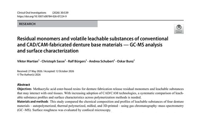
A denture can contain leftover building blocks from its manufacture. But in this study, the material with the most detectable residue did not release more of one key substance than plain control liquid.A denture resin can contain residual monomer without releasing more methyl methacrylate than a negative control. And the 3D-printed material may look chemically different for reasons a single screening method cannot fully reveal.
Reyhane Narimany, Reyhaneh Faghihian, Mehdi Jafarzadeh Samani
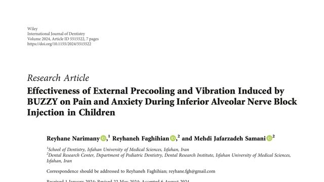
A small device combines cooling and vibration to make a dental injection less frightening for children. But when it was tested, the reassuring sensation did not clearly reduce either pain or anxiety.A device combining cold and vibration sounds like a simple way to make dental injections easier for children. But in this randomized trial, BUZZY did not significantly reduce pain or anxiety during the injection.
Bilun Jin, Heng Chen, Peiqi Liu, Yijie Wang, Yi Guo, Chenxu Wang, Yue Jia, Rui Zou, Lin Niu
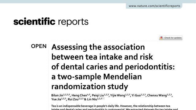
Tea is often treated as a healthy choice for your mouth. But this study found a genetic clue pointing in the opposite direction for gum disease, while finding no link with cavities.Tea is often discussed as either helpful or harmful for oral health. This study uses genetic evidence to test whether drinking more tea is actually linked to cavities, periodontitis, or tooth loss.
 3:03