A tiny change in a sugar structure may help push macrophages toward an anti-tumor state. This study traces that switch from bisecting GlcNAc, to Lgals3bp, to NF-kappa B signaling.
The functions of macrophages are governed by distinct polarization phenotypes, which can be categorized as either anti-tumor/M1 type or pro-tumor/M2 type. Glycosylation is known to play a crucial role in various cellular processes, but its influence on macrophage polarization is not well-studied. In this study, we observed a significant decrease in bisecting GlcNAc during M0-M1 polarization, and impaired bisecting GlcNAc was found to drive M0-M1 polarization. Using a glycoproteomics strategy, we identified Lgals3bp as a specific glycoprotein carrying bisecting GlcNAc. A high level of bisecting GlcNAc modification facilitated the degradation of Lgals3bp, while a low level of bisecting GlcNAc stabilized Lgals3bp. Elevated levels of Lgals3bp promoted M1 polarization through the activation of the NF-κB pathway. Conversely, the activated NF-κB pathway significantly repressed the transcription of MGAT3, leading to reduced levels of bisecting GlcNAc modification on Lgals3bp. Overall, our study highlights the impact of glycosylation on macrophage polarization and suggests the potential of engineered macrophages via glycosylated modification.
Transcript
A tiny change in a sugar structure may help push macrophages toward an anti-tumor state. This study traces that switch from bisecting GlcNAc, to Lgals3bp, to NF-kappa B signaling. Macrophages are immune cells that have emerged as promising candidates for immunotherapy in oncology, and their functions are regulated by anti-tumor M1 and pro-tumor M2 polarization phenotypes.
The M1 phenotype enhances the immune response against tumors by promoting the Th1 response, presenting antigens to cytotoxic T cells, and releasing nitric oxide and reactive oxygen species. By contrast, the M2 phenotype is characterized by anti-inflammatory cytokines, tissue repair, and angiogenesis, while nonpolarized M0 macrophages can transition into either M1 or M2 phenotypes.
That plasticity makes reprogramming M0 macrophages into the M1 phenotype an attractive pathway for immunotherapy. Recent studies have revealed that the polarization state of M1 and M2 macrophages is associated with distinct glycomic signatures. During M2 polarization, alpha two, six sialic acid and associated sialyltransferase genes are upregulated, while inhibition of sialylation in tumor-associated macrophages can reverse M2 polarization.
Elevated O-GlcNAcylation in tumor-associated macrophages has been associated with M2 polarization, whereas knockout of O-GlcNAc transferase promotes proinflammatory polarization. In cancer, increased sialylation directs macrophages toward the M2 phenotype through interaction with Siglec receptors.
The study began with an intriguing observation: bisecting GlcNAc levels decreased during M0-to-M1 macrophage polarization. That finding suggested that a deficiency in bisecting GlcNAc may contribute to driving this polarization. To investigate further, proteomics and glycoproteomics methods were used to profile differentially expressed glycoproteins and examine how proteins carrying bisecting GlcNAc modulate macrophage polarization.
Stimulation with LPS and IFN-gamma caused RAW264.7 and THP-1 macrophage cells to polarize from M0 type to M1 type. The change was evidenced by an enhanced proportion of CD86-positive cells, elevated CD86 and iNOS expression, and increased messenger RNA levels of CD86, TNF-alpha, iNOS, IL6, and IL1-beta.
Figure one establishes that LPS and interferon gamma induce M1 polarization in RAW264.7 and THP-1 macrophages, shown by increased CD86 and iNOS signals in flow cytometry and immunofluorescence. Across the same stimulation, PHA-E lectin measurements, immunofluorescence, and lectin blotting assess bisecting GlcNAc, while western blotting measures MGAT3.
The authors connect these measurements to a reduction in bisecting GlcNAc and MGAT3 during M0-to-M1 polarization. Previous studies reported higher bisecting GlcNAc levels in the M2 phenotype than in M0 or M1 phenotypes in macrophage cell lines. Here, M1 macrophages showed a significant reduction in bisecting GlcNAc compared with M0 macrophages, accompanied by downregulation of MGAT3 expression.
The decrease in bisecting GlcNAc was also observed in mouse peritoneal macrophages and bone-marrow-derived macrophages after polarization to the M1 phenotype. Silencing MGAT3 in RAW264.7 cells decreased bisecting GlcNAc levels, and that reduction was accompanied by increased CD86 expression and higher messenger RNA levels for CD86, TNF-alpha, iNOS, IL6, and IL1-beta.
The reduction in bisecting GlcNAc produced effects similar to LPS and IFN-gamma stimulation, while introducing MGAT3 hindered the M0-to-M1 polarization response after LPS and IFN-gamma treatment. Figure two tests how bisecting GlcNAc and MGAT3 affect macrophage polarization.
Silencing MGAT3 in RAW264.7 cells reduces the bisecting GlcNAc signal and is accompanied by CD86 expression in flow cytometry and immunofluorescence, while co-injection of these cells with CT26 tumor cells is associated with tumor-weight measurements and tumor-tissue macrophage staining for F4/80, PHA-E, and CD86.
Together, the panels connect altered glycosylation with macrophage activation and tumor growth in cells and mice. Because bisecting GlcNAc is a classic N-glycan structure attached to certain glycoproteins, proteomics and glycoproteomics were used to investigate proteins with differential expression and bisecting GlcNAc during RAW264.7 M0-to-M1 polarization.
The analysis identified one thousand one hundred eighteen differentially expressed proteins: five hundred eighty-three were upregulated and five hundred thirty-five were downregulated in M1 macrophages compared with M0 macrophages. Within that dataset, the analysis identified forty-two upregulated glycoproteins and twenty-five downregulated glycoproteins.
Figure three traces how the authors identified Lgals3bp as a glycoprotein carrying bisecting GlcNAc during M-zero to M-one polarization of RAW264.7 macrophages. The workflow combines proteomics and glycoproteomics, with a volcano plot, overlap diagram, and heatmap showing differential proteins and glycoproteins.
Mass spectrometry detects the modified Lgals3bp peptide ALGYEN-GlcNAc-ATQALGR, while immunoblotting and immunoprecipitation assess Lgals3bp and cellular bisecting GlcNAc. This matters because Lgals3bp expression changes alongside its glycan modification during macrophage polarization.
Using IP-MS, the study confirmed that Lgals3bp has the distinctive ion at Asp sixty-nine. Lgals3bp expression increased in the M1 type, aligning with decreased bisecting GlcNAc levels on Lgals3bp and at the whole-cellular level. Overexpression of Lgals3bp in RAW264.7 cells elevated the M1 markers CD86, TNF-alpha, and iNOS.
Conversely, silencing Lgals3bp reduced the levels of these indicative markers, and silencing Lgals3bp in RAW/shM1 cells suppressed key indicative markers of the M1 phenotype. RAW/shM1 cells, which already exhibit an M1-like phenotype, also showed increased Lgals3bp expression, supporting a role for Lgals3bp in the transition from M0 to M1 polarization.
Figure four tests whether Lgals3bp influences macrophage polarization in RAW264.7 cells. Overexpressing Lgals3bp in RAW/LG cells is accompanied by increased CD86, TNF-alpha, and iNOS expression, while silencing Lgals3bp in RAW/shLG1 cells shows the corresponding marker measurements; the figure also includes a second silencing model, RAW/shM1, and its Lgals3bp-silenced derivative.
Together, these experiments support Lgals3bp as a regulator of the M1-associated inflammatory program. RAW/shM1 cells showed less interaction between Lgals3bp and the lysosomal protein Lamp2. The N69A/LG deglycosyl mutant exhibited greater stability than WT/LG, together with reduced ubiquitination modification and less interaction with Lamp2.
MG132 or the lysosomal inhibitor chloroquine restored Lgals3bp expression, indicating that bisecting GlcNAc facilitated Lgals3bp degradation through proteasomal and lysosomal pathways. These results provide insights into how bisecting GlcNAc regulates Lgals3bp stability and its potential implications in macrophage polarization and function.
Figure five tests how bisecting GlcNAc affects Lgals3bp stability. Across immunoprecipitation, cycloheximide-chase, and ubiquitination assays, RAW cells with MGAT3 silenced show altered glycosylation, slower Lgals3bp degradation, reduced ubiquitination, and less interaction with the lysosomal protein Lamp2.
The N69A deglycosylation mutant reproduces these patterns, while MG132 and chloroquine treatments further examine proteasomal and lysosomal degradation pathways. Figure six links macrophage polarization to an NF-kappa-B–Lgals3bp–MGAT3 regulatory axis.
The GO analysis highlights NF-kappa-B, cytokine, and Toll-like receptor processes, while western blots show pathway activation after LPS and interferon-gamma stimulation or MGAT3 silencing; Co-IP also examines Lgals3bp interaction with Traf6. MGAT3 transcripts are measured during polarization, and promoter analysis predicts p65 binding, supported by dual-luciferase assays, suggesting a transcriptional connection between NF-kappa-B signaling and MGAT3.
The study emphasizes the significance of glycosylation in macrophage polarization and highlights the potential for modifying macrophages through alterations in glycosylation. The study identifies a feedback loop in which reduced bisecting GlcNAc stabilizes Lgals3bp, promotes M1 polarization through NF-kappa B, and further represses MGAT3.
That links glycosylation directly to macrophage state.
A derivative work by Paperi · AI-generated script, voice and captions
· pages and figures unaltered
Made with Paperi.
Drop in a research PDF — get a narrated video walkthrough like this one,
with highlights that follow the narration. Free to start.
Jane Callaghan, Lisa Fellin, Stavroula Mavrou, Joanne H. Alexander, Vasiliki Deligianni-Kouimtzi, Maria Papathanassiou, Judith Sixsmith
For a child living with domestic abuse, a pet may be more than a beloved animal. It may be a source of comfort, safety, and the feeling of being heard, while also becoming part of the danger.For children living with domestic violence and abuse, a pet can be much more than a pet: a family member, a source of comfort, and sometimes part of the control and danger itself.
Pratik Dutta, Matthew B. Obusan, Max Chao, Rekha Sathian, Nimisha Papineni, Ramana V. Davuluri
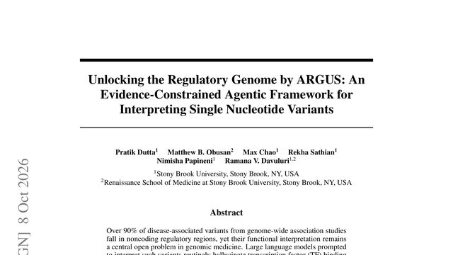
Most disease-linked changes in DNA sit outside genes, where their effects are hard to read. An AI can sound confident about them, and still invent the evidence, so this system is designed to stop and say when the evidence is not enough.What if the safest genomics AI is not the one that always gives an answer, but the one that knows when evidence cannot support a verdict? ARGUS builds that restraint directly into its investigation loop.
Mhd Hussein Murtada, Z. Faidon Brotzakis, Michele Vendruscolo
Proteins are constantly changing shape, but watching every tiny movement can take far longer than the movement itself. This study asks whether a language model can learn those motions and predict what happens under new physical conditions.What if molecular dynamics could be treated like language: structures become tokens, trajectories become sentences, and every generated path comes with an explicit probability? This paper tests that idea across proteins, temperatures, and force-field conditions.
 3:37