What if a neuron class famous for inhibiting brain activity actually helps seizures spread? This study links a remote hippocampal circuit to widespread seizures in cortical dysplasia, and identifies somatostatin-positive interneurons as an unexpected driver.
Seizures due to cortical dysplasia are notorious for their poor prognosis even with medications and surgery, likely due to the widespread seizure network. Previous studies have primarily focused on the disruption of dysplastic lesions, rather than remote regions such as the hippocampus. Here, we first quantified the epileptogenicity of the hippocampus in patients with late-stage cortical dysplasia. We further investigated the cellular substrates leading to the epileptic hippocampus, using multiscale tools including calcium imaging, optogenetics, immunohistochemistry and electrophysiology. For the first time, we revealed the role of hippocampal somatostatin-positive interneurons in cortical dysplasia-related seizures. Somatostatin-positive were recruited during cortical dysplasia-related seizures. Interestingly, optogenetic studies suggested that somatostatin-positive interneurons paradoxically facilitated seizure generalization. By contrast, parvalbumin-positive interneurons retained an inhibitory role as in controls. Electrophysiological recordings and immunohistochemical studies revealed glutamate-mediated excitatory transmission from somatostatin-positive interneurons in the dentate gyrus. Taken together, our study reveals a novel role of excitatory somatostatin-positive neurons in the seizure network and brings new insights into the cellular basis of cortical dysplasia.
Transcript
What if a neuron class famous for inhibiting brain activity actually helps seizures spread? This study links a remote hippocampal circuit to widespread seizures in cortical dysplasia—and identifies somatostatin-positive interneurons as an unexpected driver.
Seizures in cortical dysplasia tend to generalize via a large-scale seizure network, despite their localized anomalies. This is particularly a concern in late-stage cortical dysplasia, which includes focal cortical dysplasia type I, polymicrogyria, and gray matter heterotopia.
Late-stage cortical dysplasia represents one of the most common causes of pharmacoresistant epilepsy, and post-surgical relapses often arise from sources distant to the original focus. The notion of a widespread seizure network is also supported by unclear lesion margins and frequent cognitive comorbidities.
However, what drives that widespread network remains unclear. Most investigations have focused on primary lesions in the neocortex of cortical dysplasia, while the functional role of remote areas is less characterized. Abnormalities of the remote hippocampus, however, were frequently observed.
Incomplete inversion or hypoplasia of the hippocampus, as well as hippocampal hyperconnections, have been reported in patients with late-stage cortical dysplasia. Those studies left open the question of how the hippocampus, as a remote brain region, contributes to the extensive seizure network in late-stage cortical dysplasia.
The study tackled the question of a widespread seizure network in late-stage cortical dysplasia and hypothesized that hippocampal interneurons serve as a crucial gate in the network. The clinical analysis tested whether hippocampal epileptogenicity was highly associated with the widespread seizure network in late-stage cortical dysplasia.
The rat-model experiments then examined an unexpected excitatory effect of somatostatin-positive interneurons on seizure generalization, mainly attributed to neurotransmitter phenotype switching. The study included focal cortical dysplasia type I as a representative of late-stage cortical dysplasia, and type II as representing early-stage cortical dysplasia.
Fifty patients who underwent stereo-electroencephalography recordings were considered for quantitative analysis, but twelve were excluded because the epileptogenicity index could not be computed. Finally, thirty-eight patients—seventeen with type I and twenty-one with type II—were included for stereo-electroencephalography analysis, using two habitual seizures per patient.
Figure one combines SEEG mapping with clinical outcomes in focal cortical dysplasia. It shows that type one patients had more brain lobes with high epileptogenicity than type two patients, and a larger poor-prognosis group, with thirty-six percent versus seventeen percent.
Within type one, hippocampal epileptogenicity correlated with the number of regions showing high epileptogenicity, with Spearman rho equal to zero point six two and P equal to zero point zero two. Visual analysis showed that the hippocampus tended to participate in seizures of focal cortical dysplasia type I more than type II: twenty-nine point four percent versus nine point five percent in the first fifteen seconds.
Quantitative analysis found high hippocampal epileptogenicity in forty-seven point one percent of type I patients, compared with nineteen percent of type II patients. High hippocampal epileptogenicity was mainly found in patients with temporal-lobe onset, but it also appeared in extratemporal-onset type I patients more often than in type II patients.
Focal cortical dysplasia type I had a much more widespread seizure network than type II, measured by the number of brain regions with an epileptogenicity index above zero point three. Patients with type I were also more likely to have post-surgical relapses, classified as Engel class two through four, than patients with type II.
Among type I patients, a significant correlation was found between seizure-network extension and hippocampal epileptogenicity. In type II, there was no correlation with network extension or surgical outcome. To investigate the hippocampus in late-stage cortical dysplasia, the study established a rat model with prenatal freeze-lesioning at the late embryonic stage.
This model recapitulated key features of late-stage cortical dysplasia patients, including microgyria and cortical dyslamination. Figure two validates the prenatal freeze-lesion rat as a model of late-stage cortical dysplasia: staining reveals cortical dyslamination and abnormal neuronal polarity, while EEG shows interictal discharges in both cortex and hippocampus.
After low-dose PTZ, the CD rats more often developed generalized seizures and showed altered seizure timing and duration. Whole-cell recordings also indicate increased granule-cell excitability, with more evoked action potentials and a lower rheobase, linking the structural abnormalities to seizure susceptibility.
A subthreshold dose of pentylenetetrazol caused only focal seizures in most controls, with ten out of eleven controls affected that way. Remarkably, nine out of eleven cortical dysplasia rats showed generalized seizures after the same low-dose PTZ treatment, suggesting a more widespread seizure network in the model.
Seizures in cortical dysplasia rats also showed a shorter latency to generalized seizures and a prolonged generalized-seizure duration. Whole-cell recordings showed increased excitability of hippocampal dentate-gyrus granule cells, without significant differences in several intrinsic membrane properties, including resting membrane potential.
Figure three compares hippocampal structure and inhibitory interneurons in control and cortical-dysplasia rats. The widths of the dentate gyrus granule-cell layer and CA1 and CA3 pyramidal-cell layers are comparable, while NeuN-positive cell density is significantly reduced in the dentate hilus.
Fluorescence images and quantification further show a significant reduction of somatostatin-positive interneurons in the dentate gyrus, whereas parvalbumin-positive cells and interneurons in CA1 and CA3 are not marked as significantly changed. The study analyzed cell density and distribution across the dentate gyrus, CA1, and CA3.
The widths of the principal-cell layers remained unchanged between groups. NeuN-positive cell density was significantly reduced in the hilus, which includes a mix of glutamatergic cells and GABAergic interneurons, while hilar CaMKII-positive cell density was comparable between groups.
The density of parvalbumin-positive interneurons remained unchanged, but there was a dramatic reduction of somatostatin-positive interneurons in the dentate gyrus, mostly within the hilus. Somatostatin density was already reduced as early as postnatal day five, and apoptosis markers were scarcely detected from postnatal day five to day fifty, suggesting an early developmental deficit.
Figure four tracks dentate-gyrus somatostatin interneurons during PTZ-induced seizures and tests their causal role. Calcium imaging shows activity rising around generalized-seizure onset, with heatmaps indicating stage-dependent patterns; the viral labeling reached a sensitivity of seventy-seven point ninety-four plus or minus seven point seven percent and specificity of eighty-five point thirty-seven plus or minus four point seventy-eight percent.
Optogenetic inhibition and activation produced corresponding changes in seizure stage, generalized-seizure latency and duration, and progression, supporting a role for these interneurons in seizure generalization. Inhibiting somatostatin-positive interneurons significantly abolished seizure generalization, shown by fewer rats exhibiting generalized seizures, shorter generalized-seizure duration, and a rightward shift of the cumulative probability distribution.
This contrasted with a slight exacerbation of seizures when somatostatin-positive interneurons were inhibited in control rats. Activating somatostatin-positive cells in the dentate gyrus significantly accelerated seizure generalization in cortical dysplasia rats, causing prolonged generalized-seizure duration and a leftward shift of the cumulative probability distribution curve.
Initial spectral power did not change upon activation, indicating a minor role for somatostatin-positive interneurons in seizure onset. Together, the cells were increasingly recruited during generalized seizures and facilitated seizure generalization. Figure five shows that dentate-gyrus parvalbumin interneurons were weakly active during pentylenetetrazol-induced seizures, remaining hypoactive across stages.
The authors validated the calcium-imaging strategy, then used optogenetics to inhibit or activate these cells and measured seizure stage, onset latency, duration, and progression. The figure’s results support the conclusion that PV-positive interneurons attenuated cortical-dysplasia-related seizures, especially when activated.
Figure six tests whether SOM-positive interneurons directly excite dentate granule cells in control and cortical-dysplasia rats. Optogenetic stimulation and whole-cell recordings show light-evoked inhibitory and excitatory currents, while TTX with four-aminopyridine and glutamate-receptor blockers probe the synaptic mechanism.
The anatomical co-labeling of SOM, VGAT, and CaMKII supports glutamate-related marker expression in these interneurons, linking altered co-release to the excitatory recruitment of SOM cells in cortical dysplasia. Fluorescence in situ and histochemical studies examined whether GABA and glutamate can be released from individual neurons.
In cortical dysplasia rats, around thirty-three percent of somatostatin-positive interneurons co-expressed CaMKII, a marker for glutamatergic neurons, and VGAT, a marker for GABAergic neurons. In control rats, CaMKII and VGAT displayed only minimal overlap in somatostatin-positive interneurons.
Together, the results suggested an increased probability of glutamate release from somatostatin-positive interneurons in cortical dysplasia rats, producing a direct excitatory effect on downstream granule cells. Responses to most but not all stimulations suggest that only a subset of somatostatin-positive interneurons gain the co-release capability, and the signature of neurotransmitter co-release warrants further investigation with tools such as paired recordings.
Other possible mechanisms include disinhibitory connections and depolarizing GABAergic currents, but those mechanisms were considered less likely given monosynaptic transmissions and an unaltered reversal potential for GABAergic currents in cortical dysplasia rats. The findings expand the classic excitation-inhibition imbalance theory by highlighting the pro-convulsant role of somatostatin-positive interneurons, which are conventionally regarded as inhibitory, caused by neurotransmitter phenotype switching in late-stage cortical dysplasia.
The study illustrates the widespread seizure network in late-stage cortical dysplasia and provides evidence that somatostatin-positive interneurons in the dentate gyrus are crucial elements driving that large-scale network. The study proposes glutamate co-release as a novel mechanism underlying the somatostatin-positive interneuron deficits.
Further clinical evidence, including advanced neuroimaging or surgical specimens, may be needed to verify co-release in patients. Overall, hippocampal somatostatin-positive interneurons are identified as a potential therapeutic target in seizures caused by cortical malformations.
The study argues that dentate-gyrus somatostatin-positive interneurons can facilitate seizure generalization in late-stage cortical dysplasia, partly through glutamate co-release, making them a potential therapeutic target.
A derivative work by Paperi · AI-generated script, voice and captions
· pages and figures unaltered
Made with Paperi.
Drop in a research PDF — get a narrated video walkthrough like this one,
with highlights that follow the narration. Free to start.
Justin W. Andrushko, Jacob M. Levenstein, Catharina Zich, Evan Edmond, Jon Campbell, William T. Clarke, Uzay E. Emir, Jonathan P. Farthing, Charlotte Jane Stagg
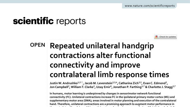
What if tiring out one hand could make the other hand respond faster? This study found that repeated squeezing with the right hand was followed by better performance in the left.What if tiring out your right hand for nine minutes could make your left hand respond faster and grip harder? This study tested that counterintuitive possibility with behaviour, functional connectivity, and brain chemistry.
Saurabh Thapliyal, Parvathi Sushama Gopinath, Dominique A. Glauser
What if hunger could turn down an animal’s heat alarm, not just making the warning quieter, but changing the alarm’s pattern so escape is no longer the default?When a worm gets hungry, it does not simply turn down its heat alarm. Starvation changes the alarm’s neural code, and flips several signals from promoting escape to suppressing it.
When you look at a group of objects, your brain can estimate their average size almost instantly. But that estimate is not fixed: another group nearby can quietly make the first group look larger or smaller.Your visual system can average a whole crowd of objects almost instantly, but those averages are not independent. Put a larger crowd beside one group, and the other group can look smaller.
 3:38