Temporal hyper-precision of brainstem neurons alters spatial sensitivity of binaural auditory processing with cochlear implants
Curious5:41CCAI
paperi.ai
0:00 / 0:00
Michaela Müller, Hongmei Hu, Mathias Dietz, Barbara Beiderbeck, Dardo N. Ferreiro, Michael Pecka
A cochlear implant can restore hearing, yet locating a sound in space can remain difficult. This study points to a surprising reason: the brain may receive timing signals that are too precise.
The ability to localize a sound source in complex environments is essential for communication and navigation. Spatial hearing relies predominantly on the comparison of differences in the arrival time of sound between the two ears, the interaural time differences (ITDs). Hearing impairments are highly detrimental to sound localization. While cochlear implants (CIs) have been successful in restoring many crucial hearing capabilities, sound localization via ITD detection with bilateral CIs remains poor. The underlying reasons are not well understood. Neuronally, ITD sensitivity is generated by coincidence detection between excitatory and inhibitory inputs from the two ears performed by specialized brainstem neurons. Due to the lack of electrophysiological brainstem recordings during CI stimulation, it is unclear to what extent the apparent deficits are caused by the binaural comparator neurons or arise already on the input level. Here, we use a bottom-up approach to compare response features between electric and acoustic stimulation in an animal model of CI hearing. Conducting extracellular single neuron recordings in gerbils, we find severe hyper-precision and moderate hyper-entrainment of both the excitatory and inhibitory brainstem inputs to the binaural comparator neurons during electrical pulse-train stimulation. This finding establishes conclusively that the binaural processing stage must cope with highly altered input statistics during CI stimulation. To estimate the consequences of these effects on ITD sensitivity, we used a computational model of the auditory brainstem. After tuning the model parameters to match its response properties to our physiological data during either stimulation type, the model predicted that ITD sensitivity to electrical pulses is maintained even for the hyper-precise inputs. However, the model exhibits severely altered spatial sensitivity during electrical stimulation compared to acoustic: while
Transcript
A cochlear implant can restore hearing, yet locating a sound in space can remain difficult. This study points to a surprising reason: the brain may receive timing signals that are too precise. Localizing sound matters for communication and navigation, but sound localization remains poor for people using two cochlear implants.
Hearing normally compares differences in when a sound reaches the two ears. The brain makes this comparison by bringing together signals from the two ears. The study found that electrical stimulation made both kinds of incoming brain signals unusually precise and somewhat more tightly locked to each pulse.
A computer model predicted that sensitivity to timing differences remained, even with these unusually precise inputs, but spatial sensitivity was severely altered compared with ordinary hearing. To find where the change began, responses to sound and electrical pulses were compared in early brain pathways in gerbils.
Neurons in both pathways showed severe hyper-precision and moderate hyper-entrainment during electrical stimulation. With unnaturally precise timing, the model preserved timing sensitivity but narrowed its useful range compared with acoustic stimulation overall.
It sharpened resolution for small timing differences, while larger differences became difficult to distinguish across the modeled range of positions. The model kept timing-difference sensitivity, but its useful range became narrower.
Small differences were easier to separate, while larger differences became indistinguishable. The biggest difference from sound was timing precision. During electrical stimulation, the timing variation was about ten times smaller.
That extreme precision continued into later brain pathways, where it was likely to influence the brain’s comparison of sounds arriving from the two ears. The model retains sensitivity to tiny left–right timing differences during electrical stimulation, but that sensitivity becomes concentrated near the centre rather than spread across the full hearing range.
Adding timing variability broadens it again, showing that input irregularity may shape where precise location judgments are possible. With ordinary sound, the model covered almost the entire physiological timing range for human hearing, with responses centered near the midline or slightly lateralized.
Its more graded response pattern also allowed the model to distinguish timing differences within either hemisphere rather than only separating left from right. With electrical stimulation, timing sensitivity remained, but it operated across a narrower range.
Responses became effectively identical for more lateralized locations on either side. The result was a strong ability to separate small differences around the middle, but almost no ability to resolve larger differences within either side—a pattern that resembles hearing left versus right without finely locating the sound.
The largest change in the inputs during electrical stimulation was the increase in response precision, meaning less timing variation. When timing variation was added back into the model to resemble sound, the useful timing range became wider, and more timing differences could be separated.
The model therefore suggests that increased precision caused by electrical stimulation directly changes how much spatial information the brain can use. The model’s extra separation near the center became weaker as more variability was added to the population response.
Because the actual variability inside a living brain was unknown, the model had to assume a particular level of that variability. However, the loss of spatial resolution at larger timing differences was mostly independent of that assumption. The experiment used animals whose hearing had fully developed before implantation, so experience-dependent tuning remained in place and degeneration had not occurred before implantation.
By contrast, people receiving implants often endure prolonged deafness, and that inactivity can degrade the auditory system and create multiple complications. The findings also suggest that some brain circuits can become functionally mature without hearing experience and may use implant-driven activity later in life to create spatial sensitivity.
Other circuits depend more strongly on hearing during an early developmental period. Among people with two implants, those whose deafness began after language learning tended to retain substantial timing sensitivity, unlike those deaf from early life.
Direct electrophysiological recordings of the cellular integration process in the LSO and MSO during implant stimulation have still not been obtained. The findings suggest that processing in one brain pathway is likely the main site where implants support timing-based spatial hearing, and that a key problem is the unusually precise timing of signals entering that comparison stage.
For people with two implants, these findings identify the unusually precise timing of incoming signals as a possible target for improving timing-based spatial hearing. The model kept left-versus-right sensitivity, but lost much of its ability to distinguish locations farther to either side.
That suggests improving the timing pattern sent into the brain could make spatial hearing more useful for people with two implants.
A derivative work by Paperi · AI-generated script, voice and captions
· pages and figures unaltered
Made with Paperi.
Drop in a research PDF — get a narrated video walkthrough like this one,
with highlights that follow the narration. Free to start.
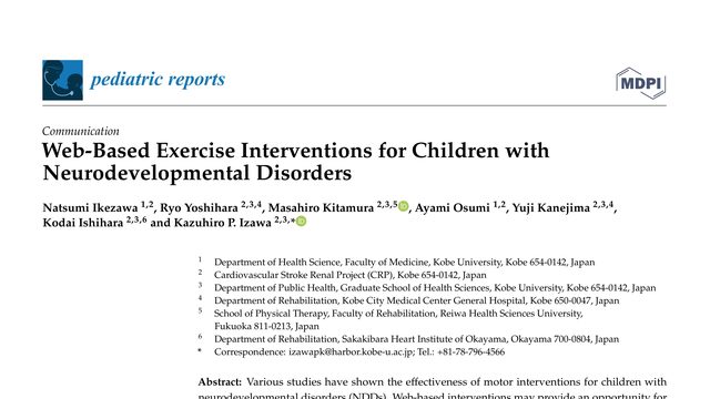
For some children, exercise support can be hard to reach. This review asks whether a screen can bring that support home—and finds encouraging results, but not for every child or every goal.What if exercise for children with neurodevelopmental disorders could happen at home, through a game console, Zoom, or WhatsApp? This review finds some encouraging improvements—but only five studies qualified.
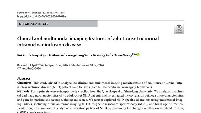
A rare brain disease can look completely different from one person to the next. But a particular pattern on a brain scan may provide a crucial clue—sometimes before doctors know what they are seeing.NIID can look like many different diseases, but a distinctive diffusion MRI pattern may reveal what is really happening. This study asks how clinical symptoms and several imaging methods fit together.
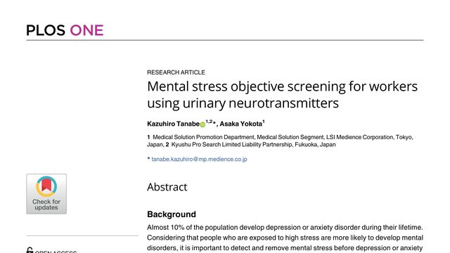
Stress can be affecting a worker before they call themselves unwell. This study asks whether a urine sample could reveal that hidden strain early enough to act on it.What if an ordinary urine sample could help reveal mental stress before it develops into depression or anxiety? This study tests that idea by combining six urinary neurotransmitters into a stress index.
 3:09
3:09