KDM3A Ablation Activates Endogenous Retrovirus Expression to Stimulate Antitumor Immunity in Gastric Cancer
Student16:20CCAI
paperi.ai
0:00 / 0:00
Jiabin Zheng, Huolun Feng, Jiatong Lin, Jianlong Zhou, Zhihui Xi, Yucheng Zhang, Fa Ling, Yongfeng Liu, Junjiang Wang, Tieying Hou, Fan Xing, Yong Li
What if disabling one epigenetic regulator could make tumor cells behave as though they were infected by a virus? This study links KDM3A removal to endogenous retroviruses, interferon signaling, and stronger immunotherapy responses in gastric cancer.
The success of immunotherapy for cancer treatment is limited by the presence of an immunosuppressive tumor microenvironment (TME); Therefore, identifying novel targets to that can reverse this immunosuppressive TME and enhance immunotherapy efficacy is essential. In this study, enrichment analysis based on publicly available single-cell and bulk RNA sequencing data from gastric cancer patients are conducted, and found that tumor-intrinsic interferon (IFN) plays a central role in TME regulation. The results shows that KDM3A over-expression suppresses the tumor-intrinsic IFN response and inhibits KDM3A, either genomically or pharmacologically, which effectively promotes IFN responses by activating endogenous retroviruses (ERVs). KDM3A ablation reconfigures the dsRNA-MAVS-IFN axis by modulating H3K4me2, enhancing the infiltration and function of CD8 T cells, and simultaneously reducing the presence of regulatory T cells, resulting in a reshaped TME in vivo. In addition, combining anti-PD1 therapy with KDM3A inhibition effectively inhibited tumor growth. In conclusions, this study highlights KDM3A as a potential target for TME remodeling and the enhancement of antitumor immunity in gastric cancer through the regulation of the ERV-MAVS-IFN axis.
Transcript
What if disabling one epigenetic regulator could make tumor cells behave as though they were infected by a virus? This study links KDM3A removal to endogenous retroviruses, interferon signaling, and stronger immunotherapy responses in gastric cancer. Immunotherapy is limited by an immunosuppressive tumor microenvironment, so identifying targets that can reverse that environment and enhance immunotherapy efficacy is essential.
The study connects tumor-intrinsic interferon with tumor-microenvironment regulation and asks whether KDM3A is suppressing that response. The proposed mechanism is that KDM3A inhibition activates endogenous retroviruses, which reshapes the double-stranded RNA, MAVS, and interferon axis and improves antitumor immunity.
Gastric cancer is common and deadly, and immunotherapy does not work equally well across tumor subtypes. Responses are mainly seen in MSI tumors, which contain more proinflammatory immune cells, while MSS tumors tend to contain more suppressed inflammatory immune cells.
That contrast motivates comparing MSI and non-MSI tumor microenvironments to find ways to reverse non-MSI immunosuppression. Epigenetic regulators may reshape the tumor microenvironment, and previous work suggests that epigenetic genes can activate tumor-intrinsic interferon through endogenous retroviruses.
ERVs generate double-stranded RNA that mimics patterns from external viruses, activating MDA5-MAVS signaling and type one interferon production. The unresolved question was how ERVs regulate the MAVS-interferon pathway in gastric cancer.
KDM3A is an epigenetic regulator that facilitates transcriptional activation by reducing histone three lysine nine dimethylation, or H3K9me2. Prior findings connect KDM3A with tumor proliferation, invasion, stemness, PD-L1 regulation, and tumor-microenvironment suppression in other cancers.
Its specific role in the gastric-cancer microenvironment remained to be investigated. The researchers analyzed a public single-cell dataset, using quality control, principal component analysis, cluster analysis, and marker genes to compare MSI and non-MSI microenvironments.
The analysis identified major cell clusters including T cells, natural killer cells, B cells, dendritic cells, and malignant cells. MSI tumors had more natural killer and T cells, higher inflammatory pathway enrichment, higher M1 macrophage scores, and lower M2 macrophage scores.
In non-MSI tumors, CD8 T cells and natural killer cells showed reduced immune activation and cytotoxicity markers. Figure one compares the tumor microenvironment in MSI and non-MSI gastric cancers, beginning with single-cell clusters and cell proportions, then examining macrophage polarization, lymphocyte expression, and T-cell receptor diversity.
The figure also shows immune-infiltration analyses in the ACRG and TCGA-STAD cohorts, with gene-set enrichment identifying the response-to-type-one-interferon pathway in the MSI subgroup. Together, these analyses support the authors’ focus on insufficient tumor-intrinsic interferon signaling as a feature of the non-MSI microenvironment.
Bulk sequencing from the ACRG and TCGA cohorts also showed more immune-activated cells in the MSI tumor microenvironment. Across both cohorts, type one interferon was significantly enriched in the MSI group. The non-MSI microenvironment therefore showed an inherent deficiency of interferon compared with MSI, potentially impairing immune activation and surveillance.
To search for an epigenetic regulator linked to interferon, the analysis correlated histone-modification genes with interferon gene sets in TCGA, GSE184336, and ACRG datasets. KDM3A was among the genes negatively associated with type one interferon gene sets and was selected from the intersection across merged datasets.
KDM3A showed a strong negative correlation with interferon gene sets. Figure two links KDM3A to suppression of tumor-intrinsic type one interferon signaling. Across datasets, the correlation analyses identify KDM3A among genes negatively associated with interferon programs, while tumor samples show elevated KDM3A expression and poorer survival, with a p-value of zero point zero one nine.
In gastric cancer cells, CRISPR disruption or IOX1 treatment increases interferon-related transcripts and IFN beta production, alongside activation of pathway proteins including phosphorylated TBK1 and IRF3, supporting KDM3A as an epigenetic brake on innate immune signaling. CRISPR-Cas9 knockout cell lines were generated in mouse MFC and human AGS gastric-cancer cell lines, and the chemical inhibitor IOX1 was also tested.
KDM3A knockout or IOX1 significantly increased CXCL10, ISG15, and IFNB1 expression in AGS and MFC cells. Knocking out KDM3A increased interferon secretion, while phosphorylation of TBK1 and IRF3 was markedly enhanced after sgKDM3A or IOX1 treatment.
RNA sequencing also showed significant enrichment of interferon-stimulated genes and interferon pathways in sgKDM3A cells. The study next asked whether endogenous retroviruses mediate the interferon response after KDM3A inhibition. In epithelial cells, type one interferon correlated positively with endogenous retroviruses, while KDM3A-positive epithelial cells correlated strongly negatively with endogenous retroviruses.
The analysis ranked LTR2B, HERVH, and LTR10C among the top three endogenous retroviruses based on prevalence. Figure three links KDM3A loss to an ERV–MAVS–interferon response. Panel A shows a negative correlation between KDM3A expression and ERV counts, with R equal to negative zero point six seven and P less than two point two times ten to the power of negative sixteen; panels B through D show ERV and cytoplasmic double-stranded RNA measurements after genetic or pharmacological KDM3A inhibition.
Panel E indicates that MAVS knockdown reduces interferon-stimulated gene expression and phosphorylated IRF3, supporting the proposed RNA-sensing pathway, while panel G examines changes in histone methylation, including H3K4me2. MAVS was knocked down in sgKDM3A AGS cells, and both interferon-stimulated-gene expression and IRF3 phosphorylation significantly decreased.
STING knockdown did not significantly affect IRF3 phosphorylation or ISG15, IFNB1, and CXCL10 expression. ERV expression did not change after MAVS or STING knockdown in sgKDM3A AGS cells. These results support type one interferon activation through double-stranded RNA signaling recognized by MAVS rather than double-stranded DNA signaling recognized by STING.
KDM3A ablation reduced gastric-cancer cell viability, and IOX1 and BIX01294 suppressed tumor growth, tumor volume, and tumor weight in BGC823 xenografts in nude mice. Transcriptome sequencing of harvested xenografts showed significant activation of endogenous-retrovirus subpopulations after KDM3A inhibition.
Figure 4 shows that pharmacological KDM3A inhibition with IOX1 or BIX01294 reduced BGC823 xenograft growth and tumor weight in nude mice. In immune-competent mice, sgKDM3A MC38 tumors also showed reduced growth and burden compared with control cells.
Crucially, anti-IFNAR1 treatment weakened this antitumor effect, supporting the authors’ conclusion that KDM3A loss suppresses tumor growth through a type one interferon-dependent mechanism. In immune-competent C57 mice, KDM3A knockout significantly inhibited MC38 tumor growth and tumor burden.
Anti-IFNAR1 antibody treatment weakened the antitumor effects of KDM3A ablation. In sgKDM3A MC38 tumors, flow cytometry found significantly greater frequencies of lymphoid cells but not myeloid cells than in control tumors.
The numbers of NK and NKT cells remained similar, while CD8 T cells increased and the proportion of CD4 T cells stayed the same. Regulatory T cells were significantly reduced in sgKDM3A tumors, and the ratio of CD8 T cells to regulatory T cells increased. CD8 T cells in sgKDM3A tumors expressed higher levels of interferon gamma and granzyme B, while PD1-positive CD8 T cells did not significantly change.
The reported immune activation involved more infiltrating immune cells, more interferon-gamma-positive CD8 T cells, and fewer regulatory T cells, independently of infiltrating myeloid cells. Figure five uses flow cytometry to compare immune cells in control and sgKDM3A MC38 tumors.
KDM3A knockout is associated with a significant increase in lymphoid cells, CD8 T cells, the CD8-to-Treg ratio, and CD8 T-cell expression of interferon gamma and Granzyme B, while NK, NKT, CD4, and PD1-positive CD8 populations show no significant change. These results indicate that KDM3A loss reshapes the tumor microenvironment toward a more activated T-cell response.
Single-cell sequencing of sorted CD45-positive cells classified six immune-cell clusters: dendritic cells, macrophages, neutrophils, natural-killer cells, B cells, and T cells. Compared with control tumors, sgKDM3A tumors had more neutrophils, natural-killer cells, and T cells but fewer macrophages.
Within the T-cell population, CD8 T cells increased, regulatory T cells decreased, and the effector-T-cell to regulatory-T-cell ratio increased. The T-cell subdivisions included multiple CD8 T-cell clusters, and sgKDM3A tumors showed a high proportion of proinflammatory T cells with decreased regulatory T cells.
Figure six uses single-cell RNA sequencing of tumor-infiltrating CD45-positive cells to map the tumor microenvironment after KDM3A ablation. The UMAP and t-SNE plots identify six major immune populations, while marker-gene heatmaps and expression maps support those cell assignments.
Bar charts then resolve immune and T-cell subtypes, including the CD8 effector-to-CD4 regulatory ratio, and violin plots compare cytotoxicity, interferon-response, and costimulatory gene expression between control and knockout tumors. The study tested whether KDM3A ablation could improve anti-PD1 therapy using KDM3A-ablated MFC and MC38 cells in combination with anti-PD1 treatment.
In C57BL/6 mice engrafted with MC38 cells, the combination of sgKDM3A and anti-PD1 significantly suppressed tumor growth compared with the control group or either single-treatment group. In MFC mouse gastrointestinal tumors treated with anti-PD1, the combination also enhanced antitumor effects compared with the control or single-treatment groups.
The findings indicate that KDM3A ablation might improve response to anti-PD1 therapy and support KDM3A as a potential immunotherapy-related target. Figure seven tests anti-PD1 therapy in two mouse tumor models: MC38 cells in C57BL/6 mice and MFC cells in six-one-five mice.
The timelines show tumor implantation followed by two intraperitoneal anti-PD1 injections, while the growth curves and tumor weights compare control and KDM3A-ablated tumors, with or without treatment. Across both models, the combination of KDM3A ablation and anti-PD1 is associated with suppressed tumor growth and reduced tumor weight, supporting KDM3A as a potential determinant of immunotherapy response.
The study identifies KDM3A as a key regulator of the non-MSI tumor microenvironment and immunotherapy sensitivity in gastric cancer. KDM3A deletion increased interferon-gamma-positive CD8 T-cell infiltration and decreased regulatory T-cell infiltration after anti-PD1 treatment in syngeneic mice.
Mechanistically, KDM3A deletion increased H3K4me2, activated endogenous-retrovirus loci, and elevated double-stranded RNA expression. The authors also observed a negative correlation between KDM3A expression and tumor-intrinsic interferon signaling or immunotherapy response in gastric-cancer patients.
A limitation is that directly knocking down endogenous retroviruses or H3K4me2 would help demonstrate their role in the MAVS-interferon axis. That experiment is technically difficult because endogenous retroviruses are numerous sequences distributed in short fragments across chromosomes, and a group rather than one specific endogenous retrovirus was upregulated.
The study also lacked specific KDM3A inhibitors and therefore used the broad-spectrum inhibitors IOX1 and BIX01294, which target the JmjC domain. Re-expression of KDM3A reversed the increases in H3K4me2, endogenous-retrovirus activation, and IRF3 phosphorylation, supporting the conclusions.
The study identifies KDM3A as a potential target for remodeling an immunosuppressive tumor microenvironment: its ablation activates the ERV-MAVS-IFN axis, improves immune-cell activity, and enhances anti-PD1 treatment in mouse tumor models.
A derivative work by Paperi · AI-generated script, voice and captions
· pages and figures unaltered
Made with Paperi.
Drop in a research PDF — get a narrated video walkthrough like this one,
with highlights that follow the narration. Free to start.
Ricky Chazya, Chilufya Mulenga, Andrew D. Gibson, Frederic Lohr, Cassandra Boutelle, Sarah Bonaparte, Oline Sinywibulula, Gareth J. Thomas, Patricia Bwalya, George Dautu, Linous Munsimbwe, Geoffrey Muuka, Luke Gamble, Ryan M. Wallace, Michelle A. Waltenburg
A rabies campaign can succeed in a rural community yet fall short in a city—not because people do not care, but because the distance to one vaccination point can decide which dogs are protected.A rabies campaign can vaccinate thousands of dogs and still miss the herd-immunity target. In Zambia, the biggest obstacle was not simply vaccine supply—it was where dogs lived, how far owners had to travel, and whether communities knew the campaign was happening.
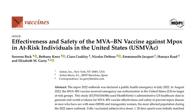
Soowoo Back, Bethany Knox, Ciara Coakley, Nicolas Deltour, Emmanuelle Jacquot, Hanaya Raad, Elizabeth M. Garry
Mpox seemed to fade from the headlines, but the risk did not disappear. In U.S. health records, people who received both recommended vaccine doses had far less mpox than similar unvaccinated people.In a small real-world US cohort, two doses of the MVA–BN vaccine were associated with eighty-nine percent effectiveness against mpox. But the same study also shows why that impressive number needs careful interpretation.
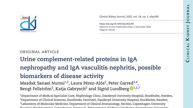
Mazdak Sanaei Nurmi, Laura Pérez‐Alós, Peter Garred, Bengt Fellström, Katja Gabrysch, Sigrid Lundberg
A kidney biopsy gives doctors a snapshot of damage, but it carries risks. This study asks whether urine can reveal something more useful: whether kidney disease is active now, or quietly leaving lasting scars.A urine sample may carry two different signals in IgA kidney disease: PTX-3 and MBL may point to active proliferative lesions, while C4c may point toward chronic damage and future decline.
 4:00
4:00