Urine complement-related proteins in IgA nephropathy and IgA vasculitis nephritis, possible biomarkers of disease activity
Student14:19CCAI
paperi.ai
0:00 / 0:00
Mazdak Sanaei Nurmi, Laura Pérez‐Alós, Peter Garred, Bengt Fellström, Katja Gabrysch, Sigrid Lundberg
A urine sample may carry two different signals in IgA kidney disease: PTX-3 and MBL may point to active proliferative lesions, while C4c may point toward chronic damage and future decline.
Introduction. The activation of the complement system plays an important role in the pathogenesis of IgA nephropathy ( IgAN) . Our primary aim was to evaluate a range of complement-related proteins, including pentraxin-3 ( PTX-3) , in blood and urine at diagnosis and their association with disease activity in the kidney biopsy, eGFR, albuminuria, and outcome. Our secondary aim was to compare the same biomarkers between patients with IgAN and IgA vasculitis with renal involvement ( IgAVN) . Methods. In a longitudinal Swedish cohort of 96 patients with IgAN ( n = 65) or IgAVN ( n = 31) , with a median follow-up time of 10.8 years, we analysed mainly lectin-pathway-related proteins and PTX-3 in plasma and urine ( u) samples stored at the time of kidney biopsy. Outcome was defined by the GFR slope or by the combined outcome of 50% loss of eGFR or end-stage kidney disease ( ESKD) . Results. Patients with detectable vs undetectable u-PTX-3 and u-mannose-binding lectin ( MBL) more frequently had mesangial hypercellularity, endocapillary proliferation, and crescents in their kidney biopsy. u-C4c levels were higher in patients with advanced tubulointerstitial fibrosis, and u-C4c was also an independent predictor of a more severe eGFR slope. There were no differences in the levels of biomarkers between patients with IgAN and IgAVN. Conclusion. u-PTX-3 and u-MBL might be biomarkers of an active proliferative stage of the disease, while higher u-C4c levels indicate more chronic lesions in both IgAN and IgAVN. These results must, however, be confirmed in larger and multiethnic cohorts.
Transcript
A urine sample may carry two different signals in IgA kidney disease: PTX-3 and MBL may point to active proliferative lesions, while C4c may point toward chronic damage and future decline. IgA nephropathy is the most common primary glomerulonephritis worldwide.
Up to 50% of patients reach end-stage kidney disease within twenty to thirty years of diagnosis, and life expectancy is reduced by six to ten years. Complement dysregulation, especially within the alternative and lectin pathways, is suggested to play key roles in the pathophysiology of the disease, leading to several ongoing phase two and three trials with complement inhibitors.
Because inflammatory activity can vary over time, non-invasive biomarkers are needed to guide individual follow-up and treatment during the disease course. IgA nephropathy is currently diagnosed exclusively from kidney biopsy findings, but biopsy does not reflect the whole individual risk profile.
Patients in Pacific Asia tend to have more progressive disease than patients in Europe, and ethnic differences have been confirmed in IgA nephropathy risk loci, including loci involved in complement regulation. Those differences highlight the importance of studying populations in different parts of the world.
Most previous complement biomarker studies were conducted in Pacific Asia, and only a few included complement analyses in urine. This leaves an important gap for larger European cohorts. Pentraxin-3 is a pattern recognition molecule in the same protein family as C-reactive protein, produced in various extrahepatic tissues and blood cells in response to inflammation and infection.
PTX-3 binds C1q, mannose-binding lectin, ficolin-1, and ficolin-2, activating complement through the classical and lectin pathways. In IgA nephropathy, mesangial cells both produce and activate PTX-3, while a previous urine PTX-3 study in lupus nephritis found increased levels in active disease.
The primary aim was to investigate PTX-3 and a wide range of circulating and urine complement-related proteins, mainly from the lectin pathway, at the time of kidney biopsy. The protein results were correlated with Oxford MEST-C histopathological findings, baseline clinical characteristics, and outcome variables.
The study also compared complement activation between IgA nephropathy and IgA vasculitis nephritis in a Scandinavian population. IgA nephropathy patients were grouped into three risk groups using the five-year risk calculated according to the international risk prediction tool for IgA nephropathy.
High risk was defined as greater than twelve percent, medium risk as four to twelve percent, and low risk as less than four percent. The final group included fourteen low-risk, twenty-five medium-risk, and twenty-six high-risk patients. Only IgA vasculitis nephritis patients with a kidney biopsy-verified diagnosis were included, and all cases with available plasma or urine samples at biopsy were enrolled.
Six IgA vasculitis nephritis biopsies could not be scored with MEST-C because the material was unrepresentative. All biopsies in the study were classified by the same single nephropathologist, and Figure 1 summarizes patient selection. Figure one traces selection from the total cohort of two hundred sixty-six patients: two hundred ten with IgAN and fifty-six with IgAVN.
After exclusions, the study retained sixty-five IgAN patients and thirty-one IgAVN patients for analysis, with the diagram also showing an expansion that added seven IgAN cases for possible MEST-C scoring. This matters because it makes the biopsy-verified study population and the reasons for sample or follow-up exclusions transparent.
C4c, C3bc, soluble C5b9, ficolins, mannose-binding lectin, collectin-11, MAP-1, MASP-2, MASP-3, and PTX-3 were quantified using in-house-developed sandwich ELISAs. For the assay, plates were coated with monoclonal antibodies and then blocked with PBS-T.
Samples and primary antibodies were diluted and incubated on the plates. HRP-conjugated reagents were added and incubated, and the plates were developed using TMB ONE as a substrate. Sulfuric acid was added to stop the reaction, and optical density was measured with a Synergy HT absorbance reader at four hundred fifty to six hundred thirty nanometers.
The same sandwich ELISAs were used for urine measurements of C4c, C3bc, soluble C5b9, ficolin-2, ficolin-3, mannose-binding lectin, MAP-1, MASP-2, MASP-3, and PTX-3, with minor modifications to adapt them for urine. Estimated glomerular filtration rate was calculated using CKD-EPI twenty twenty-one, and urine albumin measurements were converted to twenty-four-hour urinary albumin excretion to enable comparisons.
The five-year international risk prediction tool score was estimated without race according to the original formula. Outcome was defined as the average change in estimated glomerular filtration rate per year for regression analyses, or the combined outcome of fifty percent estimated filtration rate loss or end-stage kidney disease.
Mann–Whitney U tests compared medians, categorical tests compared categorical data, and Spearman rank tests evaluated correlations. Linear mixed-effects models estimated the estimated glomerular filtration rate slope, while multiple linear regression models tested whether individual biomarkers predicted albuminuria, estimated filtration rate, or filtration-rate slope.
Because many urine biomarkers had zero values, biomarkers with more than eighty-five percent zeros were excluded, and a discontinuity model was used when zero values were present. The cohort included ninety-six patients: sixty-five with IgA nephropathy and thirty-one with IgA vasculitis nephritis.
The median age was thirty-three point eight years. Median estimated glomerular filtration rate was eighty point three milliliters per minute per one point seven three square meters, median albuminuria was one point one grams per day, and the median international risk score was seven point seven percent.
Compared with IgA vasculitis nephritis, IgA nephropathy had a more severe estimated filtration rate slope during a median follow-up of ten point eight years. Table one summarizes baseline, biopsy, and follow-up characteristics for ninety-six patients: sixty-five with IgAN, thirty-one with IgAVN, and sixty controls.
At biopsy, patients had median eGFR of 80.3 milliliters per minute per 1.73 square meters and albuminuria of 1.1 grams per day; several disease–control differences were statistically significant, including creatinine, eGFR, albumin, and albuminuria. Plasma C4c, C3bc, soluble C5b9, ficolin-1, ficolin-2, and MAP-1 levels were all significantly higher in patients than in controls.
For plasma soluble C5b9, the difference was driven by patients with IgA vasculitis nephritis rather than IgA nephropathy. Table two compares complement and lectin-pathway biomarkers in plasma and urine, first between IgAN and IgAVN, and then between all disease patients and controls.
No significant plasma or urine differences were found between the two diseases; the only disease-level urine values shown are C4c and C3bc, with p-values of zero point three zero one and zero point eight three five. In contrast, several plasma markers differed between disease and control, including C4c, C3bc, sC5b9, FCN-1, FCN-2, and MAP-1, supporting disease-associated complement activation.
Because many urine values were unmeasurable, the analysis compared detectable versus undetectable biomarkers with MEST-C findings. Detectable urine MBL and PTX-3 were more frequent with M1, E1, and C1 lesions. Detectable urine soluble C5b9 was more frequent with T1, while higher urine C4c levels were detected in the presence of M1 and T1.
Table three compares detectable versus undetectable urinary biomarkers across the MEST-C components. Detectable u-MBL was associated with M1, E1, and C1, with p-values of zero point zero two two, zero point zero zero one, and zero point zero zero three, while detectable u-PTX3 showed corresponding p-values of zero point zero one one, zero point zero two zero, and zero point zero one two.
Detectable u-sC5bC9 was associated with T1, with a p-value of zero point zero three zero, illustrating how biomarker presence may reflect specific histologic features. Higher urine C4c and C3bc levels were significantly associated with both higher albuminuria and lower estimated glomerular filtration rate, even after several adjustments.
Urine ficolin-3 and urine soluble C5b9 levels were associated with the degree of albuminuria. Table 4 reports multiple linear regression coefficients for biomarker levels, stratified by detectable status, across eGFR slope, albuminuria, and eGFR in two models.
The authors highlight u-C4c and u-C3bc as associated with albuminuria and eGFR, while u-FCN3 and u-sC5b9 were associated with albuminuria; u-C4c was the only biomarker independently associated with eGFR slope after adjustment. Urine C4c was the only biomarker independently associated with a more severe estimated filtration-rate slope after adjustment for disease type, age, gender, estimated filtration rate, and albuminuria.
Median urine C4c was higher in patients who reached the combined outcome: fifty-three point two versus twenty-two point one CAU per milliliter, with a P value of zero point zero zero four. In a Cox proportional hazard model, urine C4c remained associated with the combined outcome independently of the international risk prediction score, although the hazard ratio was low: one point zero zero six.
Figure four compares urinary C4c levels in patients who did not, versus did, experience the combined outcome of ESKD or a fifty percent eGFR loss during follow-up. The distributions are shown as box plots, with individual high values visible in the group without the outcome.
The authors report median levels of 53.2 versus 22.1 CAU per milliliter, with a p-value of 0.004, supporting an association between u-C4c and the combined outcome. Figure five shows cumulative renal survival over years for patients grouped by median urinary C4c, using a Cox proportional-hazards model adjusted for the calculated IIgAN-PT risk score.
The curve for u-C4c below twenty-eight point seventy-five CAU per millilitre remains near one across follow-up, while the curve for u-C4c at least twenty-eight point seventy-five declines substantially. This supports the authors’ finding that urinary C4c was associated with the combined renal outcome even after accounting for established risk factors.
Complement-related proteins in urine might be potential biomarkers of disease activity in both IgA nephropathy and IgA vasculitis nephritis. Patients with detectable urine MBL and urine PTX-3 more frequently had proliferative histological changes. Compared with most previous European studies, the study analyzed more complement-related proteins in both plasma and urine, had a long follow-up time, and included well-characterized patients.
The cohort was relatively small and retrospective, although clinical data and samples were collected prospectively during follow-up. The measured complement proteins could not be stained in tissue, and immunosuppression during follow-up might have affected overall patient outcomes.
The number of statistical tests also increases the risk of spurious results. Urine complement-related proteins, particularly urine PTX-3, urine MBL, and urine C4c, might be potential biomarkers of disease activity and chronic changes in IgA nephropathy and IgA vasculitis nephritis.
These results must be confirmed in more extensive and prospectively designed trials. Because kidney biopsy poses risks, non-invasive biomarkers are needed to help clinicians guide follow-up and optimize treatment. The study links urinary PTX-3 and MBL with active biopsy changes, and urinary C4c with chronic lesions and worse kidney outcomes.
These markers could eventually complement, but not replace, biopsy-based assessment.
A derivative work by Paperi · AI-generated script, voice and captions
· pages and figures unaltered
Made with Paperi.
Drop in a research PDF — get a narrated video walkthrough like this one,
with highlights that follow the narration. Free to start.
Branka Bajić, Oskar Werb, Ivana Budinski, Jelena Blagojević, Juliane Schaer, Jaap van Schaik
A bat can carry a hidden infection without giving researchers an easy way to check. This study finds that the bat fly riding on its body can act like a tiny, non-invasive warning signal.What if researchers could survey bat infections without drawing blood from the bats themselves? This study shows how the bats’ own tiny, blood-feeding flies can reveal widespread and genetically diverse Polychromophilus parasites.
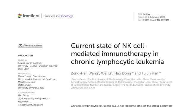
When standard leukemia treatments fall short, the body may already have another weapon: immune cells that can recognize and attack cancer without needing the same target as other immune therapies.CLL treatments have improved, but high-risk disease and drug resistance remain serious problems. This review asks whether natural killer cells can provide a more flexible way to attack leukemia.
Luke C. Cooksey, Derek C. Friesen, Enrique D. Mangan, Porunelloor A. Mathew
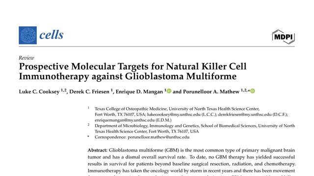
Glioblastoma is a devastating brain cancer, and standard treatment has not produced lasting survival gains. This review asks whether the body’s natural killer cells can be directed toward the tumor in several different ways.Glioblastoma is notoriously difficult to treat, but this review points to a different strategy: instead of asking NK cells to recognize one perfect tumor antigen, block the molecular brakes that keep them from attacking.
 3:37
3:37