Synthesis of L-Ornithine- and L-Glutamine-Linked PLGAs as Biodegradable Polymers
Overview4:10CCAI
paperi.ai
0:00 / 0:00
Gülce Taşkor Önel
A familiar medical plastic can be redesigned with parts borrowed from the human body, and that small change makes it become friendlier to water and break down faster. The surprising part is how useful that controlled breakdown could be.
L-ornithine and L-glutamine are amino acids used for ammonia and nitrogen transport in the human body. Novel biodegradable synthetic poly(lactic-co-glycolic acid) derivatives were synthesized via conjugation with L-ornithine or L-glutamine, which were selected due to their biological importance. L-ornithine or L-glutamine was integrated into a PLGA polymer with EDC coupling reactions as a structure developer after the synthesis of PLGA via the polycondensation and ring-opening polymerization of lactide and glycolide. The chemical, thermal, and degradation property–structure relationships of PLGA, PLGA-L-ornithine, and PLGA-L-glutamine were identified. The conjugation between PLGA and the amino acid was confirmed through observation of an increase in the number of carbonyl carbons in the range of 170–160 ppm in the 13C NMR spectrum and the signal of the amide carbonyl vibration at about 1698 cm−1 in the FTIR spectrum. The developed PLGA-L-ornithine and PLGA-L-glutamine derivatives were thermally stable and energetic materials. In addition, PLGA-L-ornithine and PLGA-L-glutamine, with their unique hydrophilic properties, had faster degradation times than PLGA in terms of surface-type erosion, which covers their requirements. L-ornithine- and L-glutamine-linked PLGAs are potential candidates for development into biodegradable PLGA-derived biopolymers that can be used as raw materials for biomaterials.
Transcript
A familiar medical plastic can be redesigned with parts borrowed from the human body—and that small change makes it become friendlier to water and break down faster. The surprising part is how useful that controlled breakdown could be. Biodegradable polymers are being developed for many medical uses and more environmentally friendly biomaterials.
Some versions have supported cell growth and helped blood vessels form. Other reported designs combined antibacterial properties with accelerated wound healing and good cell compatibility, while conductive scaffolds supported neural or cardiac tissue regeneration.
But commonly used biodegradable plastics also have drawbacks: some are expensive, break down relatively quickly, and soften at low temperatures. That limits how widely they can be used. The goal was to design and synthesize related PLGA versions by linking the polymer to amino acids as biosimilar materials.
PLGA was synthesized first from lactide and glycolide by ring-opening polymerization, and the amino acids were attached in a second step using an EDC coupling reaction. The resulting materials were checked to identify their chemical structure, size, and behavior when heated.
The amino acids were joined to PLGA by forming a strong chemical link. The two chosen amino acids were L-ornithine and L-glutamine, which were tested separately with PLGA. The researchers used PLGA as the base polymer and added amino acids through amide bonds, creating separate derivative structures with L-ornithine and L-glutamine.
The linked materials became harder to soften with heat than PLGA alone. Their heat-softening temperatures were higher, and the difference was connected with their size, structure, and ability to form more hydrogen bonds.
The higher glass-transition temperatures may reflect changes from amino-acid derivatization, with hydrogen bonding, molecular weight, and end groups contributing. The amino-acid-linked materials interacted with water more readily than PLGA.
Their water contact results supported increased water-friendliness, and both linked materials showed similar behavior. The results also reached a range associated with good cell attachment, while water-friendliness is linked with erosion beginning at the material’s surface.
The headline result came during breakdown in water: after one hundred days, PLGA had lost twenty-five percent of its weight, while the L-ornithine-linked material had lost thirty-eight percent and the L-glutamine-linked material thirty-four percent. So both amino-acid-linked materials broke down faster than PLGA.
The likely reason is that the added water-friendly groups increased the material’s chemical interaction with water. After one hundred days, the plain material had lost about a quarter of its weight, while adding either amino acid raised that loss to roughly one-third.
This matters because the added groups make the material break down faster in water. The twenty-five-day hydrolytic images showed fractures in the linked materials, indicating that breakdown had begun at their surfaces under those conditions.
The nearly straight pattern of mass loss during degradation provides additional evidence that this surface-by-surface process is surface erosion occurring. That matters for controlled drug release: a material can lose material from the outside while largely keeping its shape.
Still, body conditions or enzymes could change this surface breakdown into breakdown throughout the material. Taken together, the surface breakdown makes these materials suitable candidates for biomaterials such as controlled drug-release systems, while also warning that their behavior may change inside the body.
Linking PLGA with amino acids made it more water-friendly and faster to break down, while keeping useful heat resistance. That points toward materials that could release medicine from their surface in a more controlled way.
A derivative work by Paperi · AI-generated script, voice and captions
· pages and figures unaltered
Made with Paperi.
Drop in a research PDF — get a narrated video walkthrough like this one,
with highlights that follow the narration. Free to start.
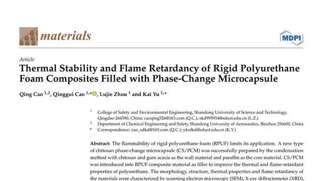
Rigid polyurethane foam is useful in cars, buildings, mining and aerospace, but it burns easily, releases toxic gases, and can trap enough heat to help start a fire. This study asks whether the same material can be made to absorb heat and slow flames.What if a material added to polyurethane foam for heat storage could also slow a fire? This study puts paraffin inside chitosan-based microcapsules and tests whether the capsules make rigid foam both thermally functional and more flame resistant.
Anastasiia Kudriavtseva, Stefan Jarić, Nikita Nekrasov, А. В. Орлов, Ivana Gadjanski, Ivan Bobrinetskiy, Petr I. Nikitin, Nikola Ž. Knežević
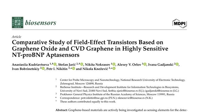
Could a saliva sample reveal signs of heart failure without a blood draw? This study found that a less perfect form of graphene could detect the signal more reliably than the cleaner, more orderly version.Two graphene-based sensors cost about the same, but they do not perform the same: the rougher, more defective material detects NT-proBNP in artificial saliva at a lower concentration than pristine CVD graphene.
Hugo X. Rodrigues, Hudson R. Armando, Daniel A. da Silva, João Paulo C. L. da Costa, Luiz Antônio Ribeiro, Marcelo Lopes Pereira
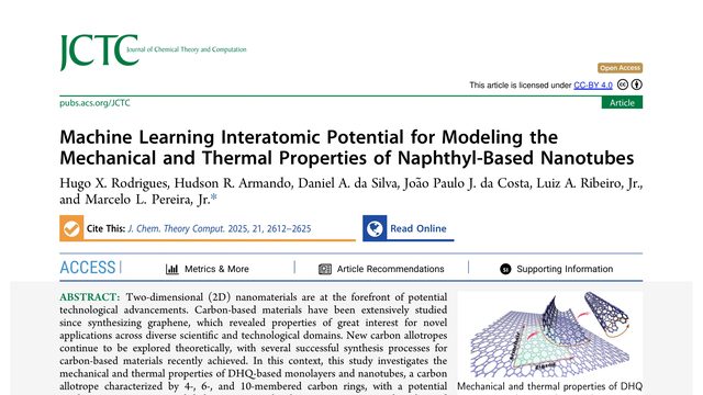
A tiny carbon structure can stay intact at temperatures far beyond ordinary industrial heat, yet snap suddenly when stretched. This study builds a faster way to find out exactly where those limits lie.What happens when a carbon nanomaterial is pushed, stretched, and heated far beyond ordinary conditions? This study uses a machine-learned interatomic potential to test DHQ sheets and nanotubes at scales first-principles calculations cannot reach.
 4:54
4:54