Single-molecule visualization of mRNA circularization during translation
Overview4:22CCAI
paperi.ai
0:00 / 0:00
Byungju Kim, Jincheol Seol, Yoon Ki Kim, Jong‐Bong Lee
Inside every living cell, tiny messages are read to build proteins. Scientists thought many of those messages formed helpful loops, but watching them one at a time suggests the loop may be an illusion of motion.
Translation is mediated by precisely orchestrated sequential interactions among translation initiation components, mRNA, and ribosomes. Biochemical, structural, and genetic techniques have revealed the fundamental mechanism that determines what occurs and when, where and in what order. Most mRNAs are circularized via the eIF4E–eIF4G–PABP interaction, which stabilizes mRNAs and enhances translation by recycling ribosomes. However, studies using single-molecule fluorescence imaging have allowed for the visualization of complex data that opposes the traditional “functional circularization” theory. Here, we briefly introduce single-molecule techniques applied to studies on mRNA circularization and describe the results of in vitro and live-cell imaging. Finally, we discuss relevant insights and questions gained from single-molecule research related to translation.
Transcript
Inside every living cell, tiny messages are read to build proteins. Scientists thought many of those messages formed helpful loops—but watching them one at a time suggests the loop may be an illusion of motion. Protein production proceeds through precisely orchestrated, sequential interactions among translation initiation components, messenger RNA, and ribosomes, with mechanisms determining what occurs, when, where, and in what order.
Most messenger RNAs form a circular loop through eIF4E, eIF4G, and PABP; this stabilizes them and helps recycle ribosomes to enhance translation. Watching individual messages with single-molecule fluorescence imaging has produced complex data that opposes the traditional theory of functional circularization during translation.
The usual picture is like a reusable instruction sheet whose two ends are clipped together: the clip could protect the sheet and help the reader return quickly to the beginning. In cells, the two ends can communicate through binding proteins, but those interactions are random rather than fixed.
The loop can change dynamically, so its shape during reading has remained poorly understood. To follow that changing shape, the researchers use light from two attached glowing markers. When the markers come within several nanometers, energy moves between them, and the changing signal reveals how close they are in real time.
This approach tracks interactions between molecules or distances between residues by measuring energy transfer efficiency from a donor fluorophore to an acceptor. In purified systems, eIF4G increases eIF4E’s affinity for the message’s five-prime cap, strengthening their attraction at the front end.
Pab1p, a yeast polyadenylate-binding protein, has an even stronger effect, bringing both RNA ends together even without polyadenylate or eIF4G. The observation suggests that this helper can change the message’s shape by touching the message directly, although its interaction with the message itself is weaker than its interaction with the tail.
Messenger RNA naturally folds so its two ends stay close, but translation pushes those ends farther apart as more ribosomes occupy it. This makes end-to-end distance a visible readout of how actively the message is being translated and whether translation begins at its capped end or an internal entry site.
In cells, the proteins expected to connect the two RNA ends rarely appear together, with the experiments finding rare colocalization between these factors. Most messenger RNAs contain only one member of the tested set of connecting factors, rather than showing both members together in the same message.
Under normal conditions, that result suggests that a message may not be physically circularized by those proteins while translation is active. A modified message can improve translation without increasing this end-to-end connection.
The findings raise a puzzle: how can ribosomes reinitiate after termination, and does communication between both messenger RNA ends occur only in a nontranslating state? Another possibility is that the first ribosome carries a connecting helper toward the far end, creating the physical link only after that ribosome finishes.
These possibilities remain open questions. The old picture treated protein links as the standard explanation for message circularization. The newer observations show that a message can look circularized during active translation even when proteins do not physically connect its ends.
The practical lesson is that scientists should follow individual messenger ribonucleoprotein complexes inside living cells, using single-molecule fluorescence imaging directly. That could reveal how changing interactions alter messenger RNA conformation throughout translation and how those interactions might eventually be changed.
The message can look compact while it is being read, without staying physically tied at both ends. That changes how scientists think about protein production and what they must measure next in living cells.
A derivative work by Paperi · AI-generated script, voice and captions
· pages and figures unaltered
Made with Paperi.
Drop in a research PDF — get a narrated video walkthrough like this one,
with highlights that follow the narration. Free to start.
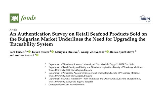
Lara Tinacci, Deyan Stratev, Mariyana Strateva, G. Zhelyazkov, Ralica Kyuchukova, Andrea Armani
A seafood label can promise one species while the food inside is another. In Bulgaria, a broad check of shop-bought seafood found that this was not a rare mistake, but a problem affecting about one product in nine.A seafood label can look perfectly ordinary while naming the wrong species. In Bulgaria, DNA testing found that eleven percent of the sampled products were mislabeled, exposing both consumer risks and weaknesses in traceability.
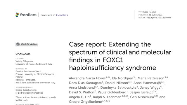
Alexandra Garza Flores, I. Nordgren, Maria Pettersson, Dora Dias‐Santagata, Daniel Nilsson, Anna Hammarsjö, Anna Lindstrand, Dominyka Batkovskyte, Janey L. Wiggs, David S. Walton, Paula Goldenberg, Jesper Eisfeldt, Angela E. Lin, Ralph S. Lachman, Gen Nishimura, Giedré Grigelioniené
A condition often recognized through the eyes can reach far beyond vision. In two adults, the same missing genetic instruction was linked to glaucoma, unusual bones, loose joints, and problems that became clearer with age.What happens when losing one copy of a developmental gene produces not just eye disease, but a much broader skeletal and neurologic picture? This case report shows how genome sequencing exposed that expanded spectrum.
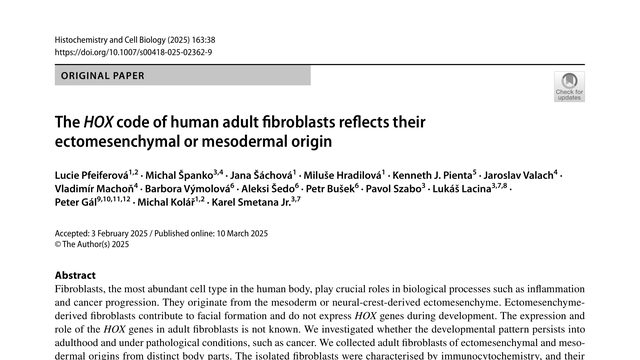
Lucie Pfeiferová, Michal Španko, Jana Šáchová, Miluše Hradilová, Kenneth J. Pienta, Jaroslav Valach, Vladimír Machoň, B Vymolová, Aleksi Šedo, Petr Bušek, Pavol Szabó, Lukáš Lacina, Péter Gál, Michal Kolář, Karel Smetana
Two fibroblasts can look almost identical under a microscope, yet one may still carry a molecular record of the body region it came from, and that record can remain visible in cancer.Two adult fibroblast populations can look almost identical under the microscope yet carry strikingly different developmental signatures. This study asks whether HOX genes preserve a cell’s embryonic address long after development ends.
 4:33
4:33