Case report: Extending the spectrum of clinical and molecular findings in FOXC1 haploinsufficiency syndrome
Overview4:30CCAI
paperi.ai
0:00 / 0:00
Alexandra Garza Flores, I. Nordgren, Maria Pettersson, Dora Dias‐Santagata, Daniel Nilsson, Anna Hammarsjö, Anna Lindstrand, Dominyka Batkovskyte, Janey L. Wiggs, David S. Walton, Paula Goldenberg, Jesper Eisfeldt, Angela E. Lin, Ralph S. Lachman, Gen Nishimura, Giedré Grigelioniené
A condition often recognized through the eyes can reach far beyond vision. In two adults, the same missing genetic instruction was linked to glaucoma, unusual bones, loose joints, and problems that became clearer with age.
A condition often recognized through the eyes can reach far beyond vision. In two adults, the same missing genetic instruction was linked to glaucoma, unusual bones, loose joints, and problems that became clearer with age.
FOXC1 is a genetic instruction that plays a critical role during early development. When harmful changes affect it, people can develop eye abnormalities with a high risk of glaucoma, along with changes in the face, teeth, bones, hearing, and heart.
Here is the everyday picture: imagine a building plan used during construction. If one important instruction is missing, the effects may appear in several parts of the building, not just one room. That is the pattern described here.
The report describes two unrelated adult women with reduced FOXC1 activity. Both had eye and skeletal abnormalities, and both received a genetic diagnosis through genome sequencing. The eye condition linked to FOXC1 is Axenfeld-Rieger syndrome, a condition involving the front part of the eye and a high risk of glaucoma and cataracts.
It can also include distinctive facial features, unusual teeth, heart and kidney problems, joint pain, and short stature. The range is wider than these familiar features. Less common findings include changes in the brain, such as enlarged fluid spaces and underdevelopment of a part of the brain involved in coordination.
FOXC1-related changes can take several forms, including changes that alter the gene's message, missing stretches, or extra copies. More than four hundred people with certain disease-causing FOXC1 changes had already been reported. The two women had overlapping features caused by reduced FOXC1 activity.
One had a complex rearrangement in the region containing FOXC1; the other had a change that produced an early stop in the same gene. But the story was not limited to the eyes. Both women had significant skeletal abnormalities, and both needed orthopedic surgery.
The first woman's genetic change was especially complicated: a missing piece containing FOXC1 was followed by a very large section turned around, and then another missing piece. The complex rearrangement included a missing piece containing FOXC1, followed by a large section turned around, and then another missing piece.
Across childhood and adulthood, both people retain a recognizable facial appearance alongside unusually shaped bones, spinal problems, and hip or elbow abnormalities—showing that this condition affects the whole body and persists over time. The complex rearrangement may help explain why the first woman had a more severe condition.
The large inverted section includes several genes important for development. The genome is not just a line of instructions; its three-dimensional structure helps control which instructions are used. The inversion may have disturbed that structure across many genes, contributing to the more severe condition.
Reduced FOXC1 activity is associated with a spectrum, from isolated anterior segment dysgenesis through Axenfeld-Rieger syndrome to a phenotype overlapping De Hauwere syndrome. The skeletal manifestations are likely under-ascertained and may become more apparent as the patient ages, so awareness should increase over time.
People with these eye findings should also be screened for skeletal problems, with a low threshold for obtaining a skeletal survey. A broader multisystem evaluation should also consider the brain, hearing, heart, teeth, and orthopedic health, rather than focusing only on the eyes.
For patients and families, a multisystem approach matters even when anterior segment dysgenesis appears isolated at first, because FOXC1-related anomalies can extend beyond the eyes. Increased awareness of these specific risks can support more personalized follow-up, even though the right follow-up timing is difficult because phenotype varies.
The key message is practical: reduced FOXC1 activity can affect several body systems, and hidden skeletal problems may be missed unless care looks beyond the eyes. Earlier, broader checks could make care more personal.
A derivative work by Paperi · AI-generated script, voice and captions
· pages and figures unaltered
Made with Paperi.
Drop in a research PDF — get a narrated video walkthrough like this one,
with highlights that follow the narration. Free to start.
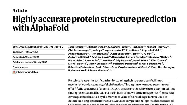
Lucie Pfeiferová, Michal Španko, Jana Šáchová, Miluše Hradilová, Kenneth J. Pienta, Jaroslav Valach, Vladimír Machoň, B Vymolová, Aleksi Šedo, Petr Bušek, Pavol Szabó, Lukáš Lacina, Péter Gál, Michal Kolář, Karel Smetana
Two fibroblasts can look almost identical under a microscope, yet one may still carry a molecular record of the body region it came from, and that record can remain visible in cancer.Two adult fibroblast populations can look almost identical under the microscope yet carry strikingly different developmental signatures. This study asks whether HOX genes preserve a cell’s embryonic address long after development ends.
As tea leaves grow older, they make less caffeine, and this study points to a biological switch that may connect caffeine loss with the leaf’s aging process.What if a tea plant’s aging program also controls how much caffeine its leaves contain? This study identifies CsS40, a senescence-related transcription factor linked to lower TCS1 expression and reduced caffeine accumulation.
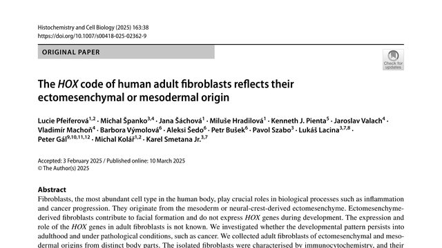
John Jumper, Richard Evans, Alexander Pritzel, Tim Green, Michael Figurnov, Olaf Ronneberger, Kathryn Tunyasuvunakool, Russ Bates, Augustin Žídek, Anna Potapenko, Alex Bridgland, Clemens Meyer, Simon A. A. Kohl, Andrew J. Ballard, Andrew Cowie, Bernardino Romera-Paredes, Stanislav Nikolov, Rishub Jain, Jonas Adler, Trevor Back, Stig Petersen, David Reiman, Ellen Clancy, Michał Zieliński, Martin Steinegger, Michalina Pacholska, Tamas Berghammer, Sebastian Bodenstein, David Silver, Oriol Vinyals, Andrew W. Senior, Koray Kavukcuoglu, Pushmeet Kohli, Demis Hassabis
Every cell depends on proteins, but scientists have seen the shape of only a tiny fraction of them. AlphaFold showed that a sequence of chemical letters can be enough to predict a protein’s three-dimensional form with near-experimental accuracy.For more than fifty years, predicting a protein’s three-dimensional shape from its sequence remained an open problem. AlphaFold changed the scale of what was possible, reaching near experimental accuracy in many cases, even without a similar known structure.
 4:36
4:36